Pathophysiology · Digestive, renal, and perfusion
Volume loss: digestive and renal convergence, divergence toward organ perfusion
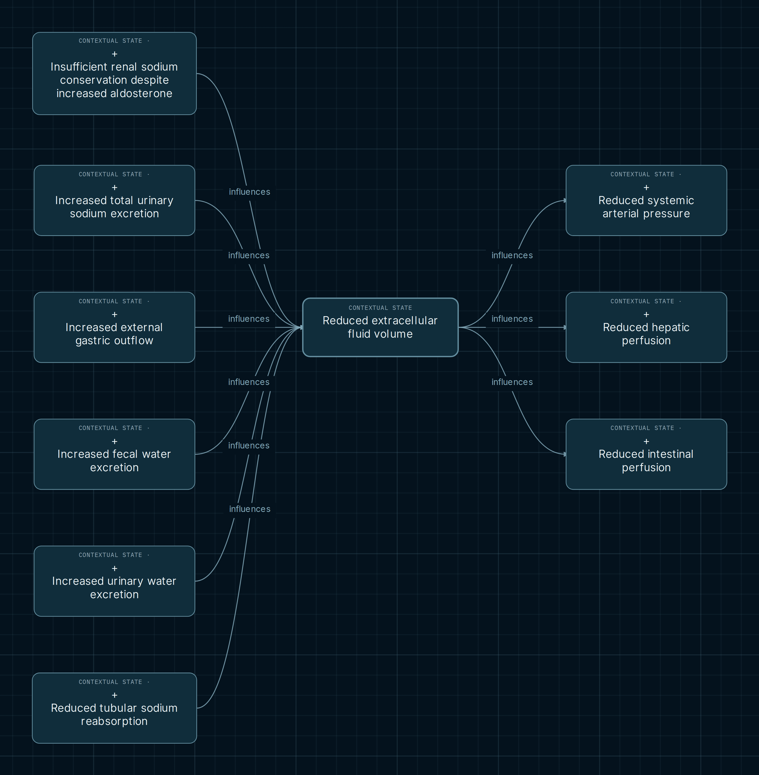
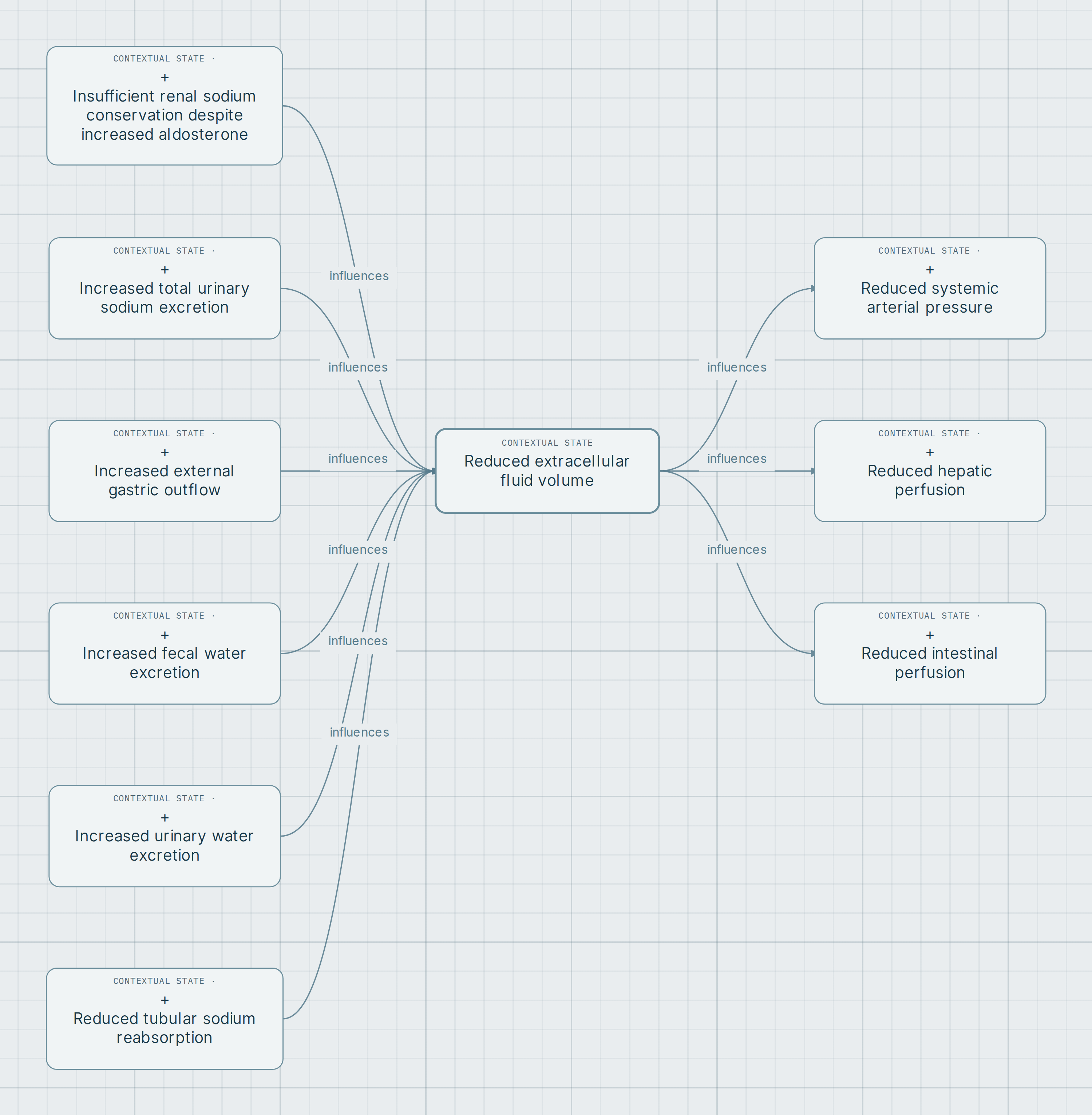
Extracellular volume brings together processes often studied in separate chapters. Six digestive and renal inputs converge in this selection, while three outputs allow comparison of arterial pressure and regional perfusion. Reading these branches together identifies a shared bodily state without assuming that all losses have the same composition or produce the same outcome.
Explore this map in the AtlasOpens chains from reduced extracellular fluid volume, in both directions and at depth 1.
The map
Reading balance and influence
The center describes a bodily compartment; placement in the urinary system does not make it a volume located inside the kidney. Each input retains balance and compensation conditions. The three outputs are separate influences from the shared node: this view does not draw an obligatory chain from arterial pressure to liver and then intestine.
- Loss and balance
- An increased output per unit time must be compared with intake, other losses, composition, and the interval examined. It does not establish net bodily loss by itself.
- Conditional influence
- Each arrow links states under explicit conditions. Notes about the general physiological relationship may differ in scope from notes about its contextual application.
- Compartments and perfusion
- Extracellular volume, circulating volume, arterial pressure, and regional flow are different variables. Their convergence or divergence in the map does not make them equivalent.
Volume inputs and outputs
Two digestive inputs
Increased fecal water excretion requires unreplaced net external loss, considering solutes, redistribution, and renal compensation. External gastric outflow represents documented vomiting or drainage; nausea or luminal secretion subsequently reabsorbed does not equal that outflow. Both pathways share a destination while retaining their composition and context.
Four renal inputs
The inputs are increased urinary water excretion, increased total urinary sodium excretion, reduced tubular sodium reabsorption, and insufficient renal sodium conservation despite increased aldosterone. They are not interchangeable measurements: distinguish total flow, concentration, filtered load, and distal response before attributing contraction to an isolated sample.
Three hemodynamic outputs
Lower arterial pressure requires impaired filling or effective circulation with insufficient compensation. Hepatic perfusion requires assessment of dual inflow and arterial compensation; intestinal perfusion requires territory, distribution, outflow pressure, and demand. Volume reduction alone does not demonstrate ischemia in either territory.
Excretion, reabsorption, and hormonal response
Urinary water excretion is volume per time, not voiding frequency. Total sodium excretion is neither urinary sodium concentration nor fractional excretion. NIDDK and OpenStax teaching material separates filtration, tubular recovery, and final elimination; the map notes add conditions for whole-body balance.
Low absolute reabsorption with low filtration does not demonstrate net salt loss. The branch with elevated aldosterone requires sufficient bioactive exposure and insufficient distal conservation, with other causes evaluated. Its name describes a joint functional configuration; it does not establish a genetic disorder or a hormonal etiology.
Compare territories without adding effects
The three outgoing arrows do not justify adding reductions together or assigning a temporal sequence. A regional response depends on flow distribution and compensation. The WSES guideline provides context for the intestinal branch, but its diagnostic categories and treatment recommendations do not become nodes or instructions in this selection.
Depth 1 shows immediate neighbors. It does not demonstrate the absence of intermediate mechanisms or include all renal and digestive regulation. The useful academic exercise is to identify the additional condition needed to move from external loss to documented contraction, and from contraction to a particular regional effect.
Unreplaced loss connects organs; an isolated value does not reconstruct balance
To compare a digestive branch with a renal branch, retain the same interval and specify intake, output, and redistribution. Then examine each regional output separately. This integrates systems without deriving extracellular volume from serum sodium concentration or regional perfusion from systemic pressure alone.
Explore the differences
- Locate the shared volume node and compare the two digestive inputs with the four renal inputs. Identify where unreplaced net loss is required.
- Open “Notes and legend” for sodium excretion and tubular reabsorption. Separate the general physiological relationship from its conditions in these states.
- Examine the three outgoing connections separately. Compare circulatory compensation, dual hepatic supply, and intestinal perfusion conditions.
Opens chains from reduced extracellular fluid volume, in both directions and at depth 1.
Scope and sources
States retain editorial-proposal scope; connections combine editorial proposal and partial support, while some underlying relationship notes say “source verified”. No label automatically validates the whole network. There is no diagnosis of shock or ischemia, universal threshold, or inevitable progression. This is a draft for academic review.
References recorded in the notes. Sources explain particular functions or contexts; graph scope remains conditional and does not adopt their clinical protocols:
- NIDDK / NIH. Your Kidneys & How They Work. Filtration, tubular recovery, and water and salt balance.
- OpenStax / Rice University. Anatomy and Physiology 2e, 25.6: Tubular Reabsorption.
- OpenStax / Rice University. Anatomy and Physiology 2e, 23.7: Chemical Digestion and Absorption. Locators: water and electrolyte absorption.
- Bala M et al. Acute mesenteric ischemia: updated guidelines of the World Society of Emergency Surgery. 2022;17:54. DOI: 10.1186/s13017-022-00443-x. Context for the intestinal branch.
- AJKD. Metabolic Alkalosis Pathogenesis, Diagnosis, and Treatment: Core Curriculum 2022. Note reference for gastric losses and renal responses.
- Merck Manual Professional. Overview of Disorders of Fluid Volume. Reference for the extracellular volume state and sodium/water balance.
- Clinical Methods, Jaundice. Basic Science and Clinical Significance. Recorded reference for reduced hepatic perfusion; not quantitative validation of this branch.