Pathophysiology · Cardiovascular and pulmonary circulation
Pulmonary congestion: converging pressures and diverging flows
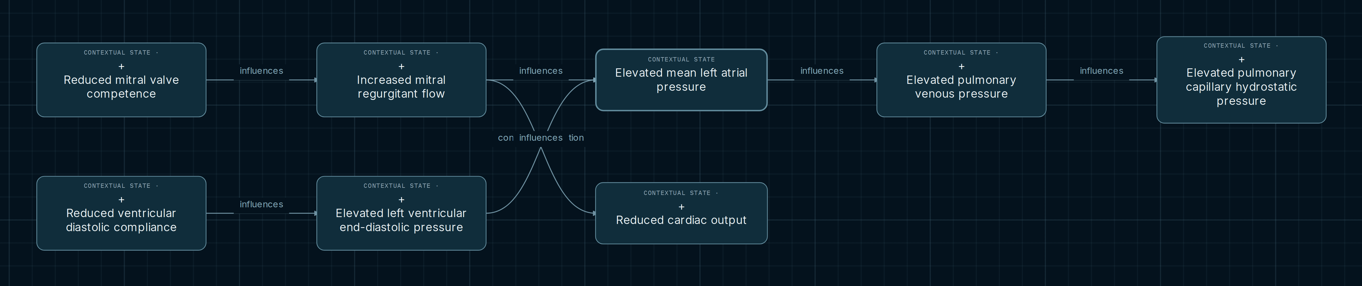
Elevated left atrial pressure can occur in different functional contexts. This selection compares a ventricular branch with a mitral branch, identifies their convergence, and follows a divergence between pulmonary pressure and systemic flow. The aim is to read conditional relationships while preserving distinctions between chambers, cardiac phases, and measurement methods.
Explore this map in the AtlasOpens chains from elevated mean left atrial pressure, in both directions and at depth 2.
The map
Reading the connections
Start at the selected node and compare incoming arrows before following outgoing ones. Visual proximity does not express severity or duration. Depth 2 bounds the selection; it does not establish the number of stages in a biological process. The only transition connects elevated left ventricular end-diastolic pressure to elevated mean left atrial pressure.
- Contextual influence
- Links applications of variables under recorded conditions. The arrow does not calculate magnitude, probability, or the net effect of combining several states.
- Editorial transition
- The ventricular–atrial connection proposes a conditional reading of filling. It is not a causal influence and does not turn two different pressures into a shared measurement.
- Convergence and divergence
- Two inputs share the atrial pressure node. Regurgitant flow also connects to reduced cardiac output: a shared origin does not require both destinations to occur.
Two inputs and two destinations
Ventricular branch
Lower diastolic compliance relates to higher end-diastolic pressure when volume, external pressure, and the relevant region of a documented curve are comparable. An isolated slope cannot establish absolute pressure. The subsequent transition requires consideration of filling gradients, the mitral valve, and loading.
Mitral branch
Reduced mitral competence permits backward flow through a communicating orifice under a ventricular–atrial gradient during systole. Linking this flow to higher mean atrial pressure requires effective loading, comparable compliance and emptying, and insufficient compensation. A v-wave peak alone does not establish an elevated mean.
Pulmonary pressures and output
Atrial pressure connects to pulmonary venous pressure, which connects to capillary hydrostatic pressure. Circuit continuity and comparable flow, resistance, and loading are required. The reduced-output branch requires comparable total ejection and heart rate with insufficient compensation; backward flow differs from net systemic flow.
Four pressure names, four different questions
Ventricular end-diastolic pressure describes one instant; mean atrial pressure summarizes a cycle in another chamber. Venous pressure belongs to a vascular territory, and capillary hydrostatic pressure to pulmonary microcirculation. Keeping these definitions distinct prevents substitution of one variable for another when a chain looks continuous.
Wedge pressure may approximate atrial pressure under technical and physiological assumptions, but it does not directly measure every capillary. Reddy and colleagues combine invasive hemodynamics with lung ultrasound during exercise, providing relevant human evidence without making these measurements universally interchangeable.
Where this selection stops
The title frames a question about congestion; the captured selection stops before representing pulmonary fluid accumulation. Following arrows to capillary pressure therefore does not establish edema in an individual. The HFA/ESC review supplies context about congestion, while the graph retains its particular states and conditions.
Also compare passive stiffness with active relaxation: the compliance note requires that distinction and does not assume fibrosis. Likewise, reduced mitral competence describes contextual function without requiring a particular structural lesion. These distinctions support studying mechanisms without treating every node as a disease.
Convergence on a pressure does not identify its cause
Finding the shared node does not justify tracing an arrow backward to select an origin. Elevated pulmonary venous pressure also does not prove elevated atrial pressure: the notes allow for regional venous obstruction. Use the map to formulate comparisons and examine conditions, rather than infer etiology from its shape.
Explore the conditions
- Open the selection and locate the shared atrial pressure. Follow the ventricular and mitral inputs separately.
- Select the ventricular–atrial transition and examine its conditions in “Notes and legend”. Compare it with an influence.
- Follow both outgoing connections from regurgitant flow. Identify conditions allowing cardiac output to remain preserved and those conditioning atrial pressure.
Opens chains from elevated mean left atrial pressure, in both directions and at depth 2.
Scope and sources
States and connections in this selection retain “editorial proposal” support status. References support concepts and contexts; they do not validate the entire network as a predictive model. There are no diagnostic thresholds, universal timelines, or inevitable progression. This guide is a draft for academic review.
Actual references from Atlas notes. Distinguish the human study, institutional review, and physiology teaching sources when assessing the support for each connection:
- Reddy YNV et al. The haemodynamic basis of lung congestion during exercise in heart failure with preserved ejection fraction. European Heart Journal. 2019;40:3721–3730. DOI: 10.1093/eurheartj/ehz713.
- Harjola VP et al., HFA/ESC. Organ dysfunction, injury and failure in acute heart failure. European Journal of Heart Failure. 2017;19:821–836. DOI: 10.1002/ejhf.872. Congestion section and figure 1.
- OpenStax / Rice University. Anatomy and Physiology 2e, 19.3: Cardiac Cycle. Note locator: Pressures and Flow.
- CV Physiology. Valvular Insufficiency. Recorded reference for gradients, mitral regurgitation, and total ejection versus forward flow.
- OpenStax / Rice University. Anatomy and Physiology 2e, 20.2: Blood Flow, Blood Pressure, and Resistance. Venous pressure relationships.