Physiology · Nervous and musculoskeletal systems
Patellar and Achilles reflexes: a shared sequence, different circuits
A tendon tap may produce a brief response, but sensory reception, spinal integration, neuromuscular transmission and contraction lie between the stimulus and muscle tension. Comparing the patellar and Achilles reflexes reveals the organization they share and what changes when it is placed in different muscles, nerves and spinal segments.
Explore this map in the AtlasOpens both reflexes together. Click a component to expand its anatomical sites; the notes separate each circuit’s applications.
Two views of the circuit
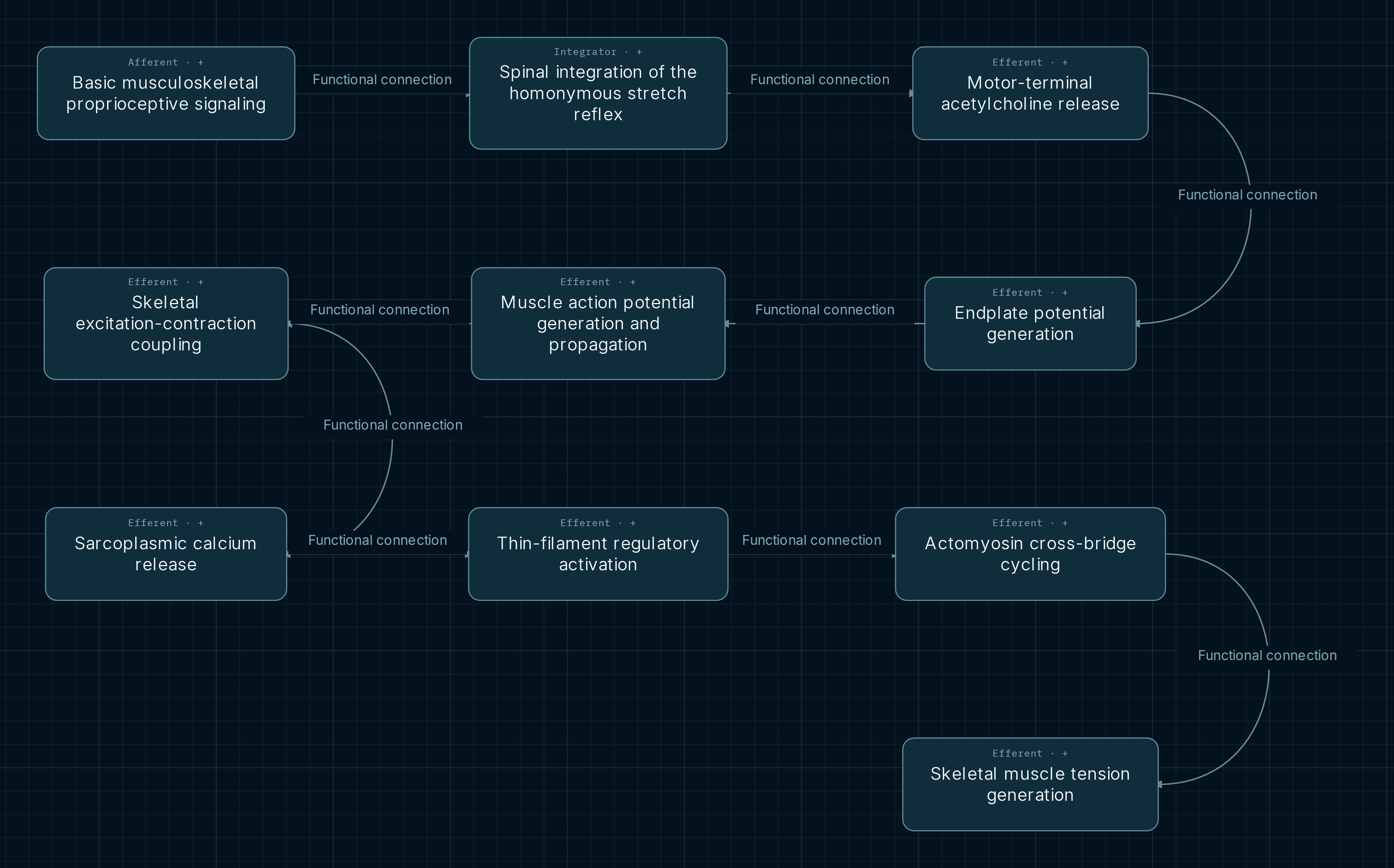
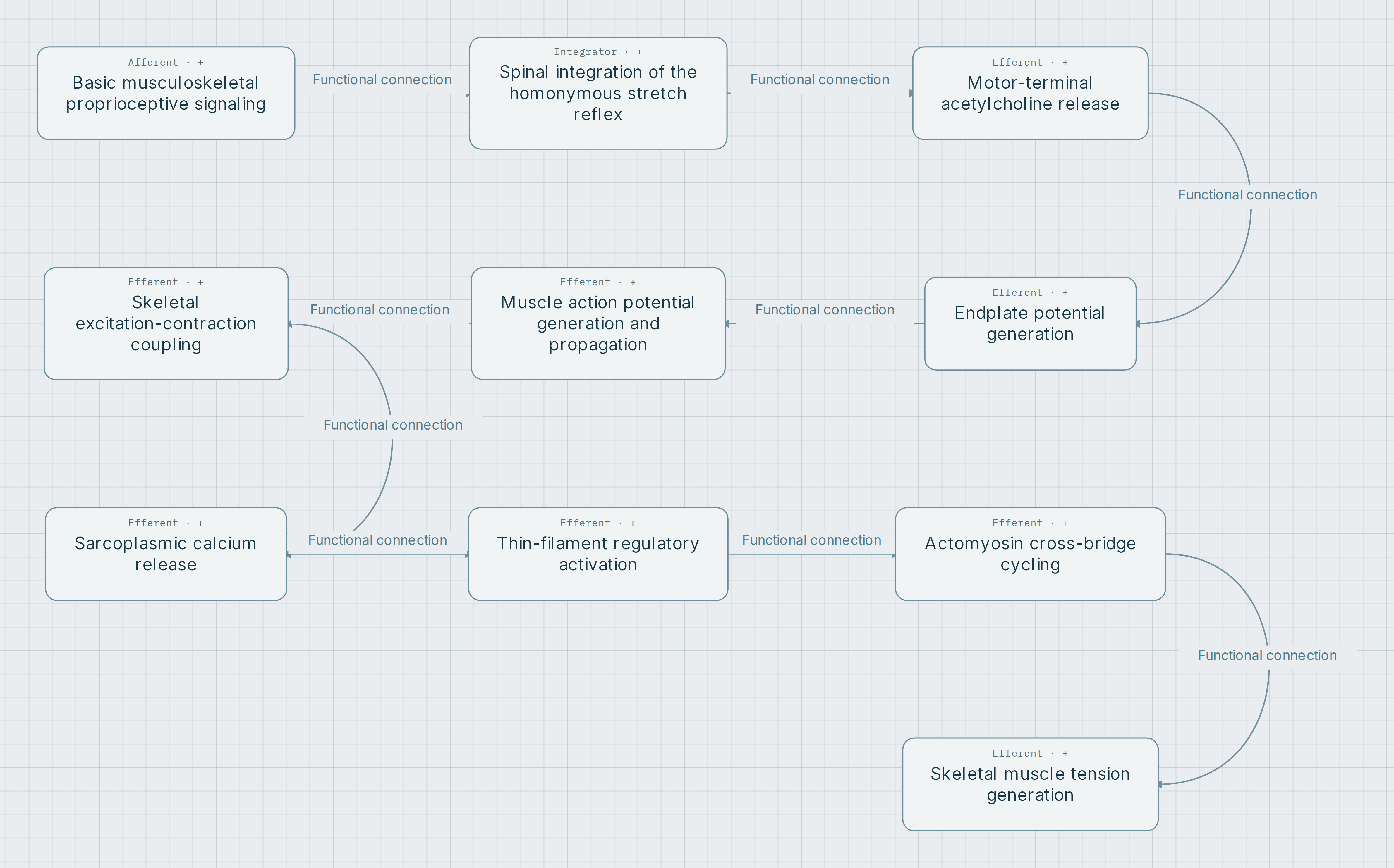
Where each circuit takes place
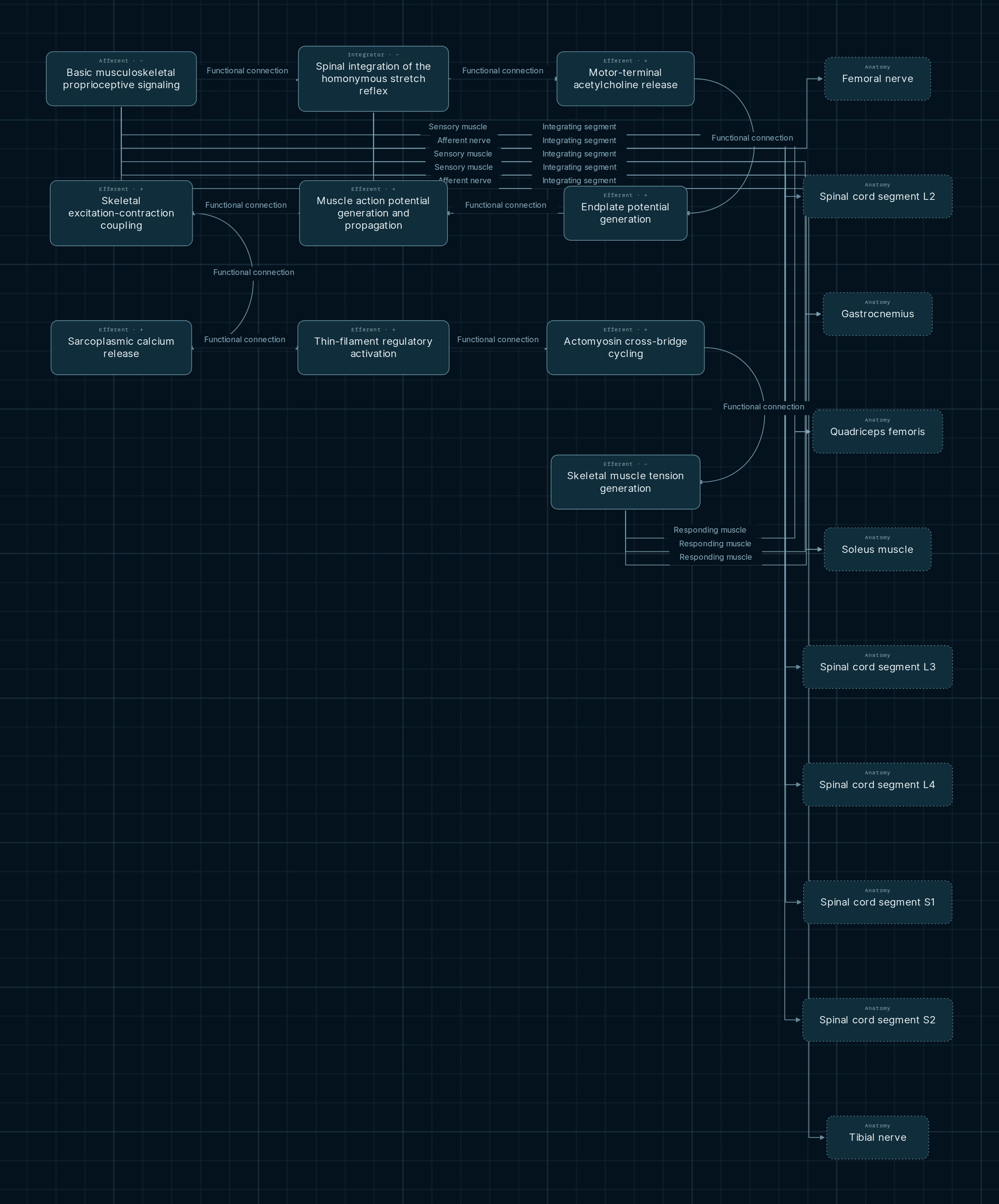
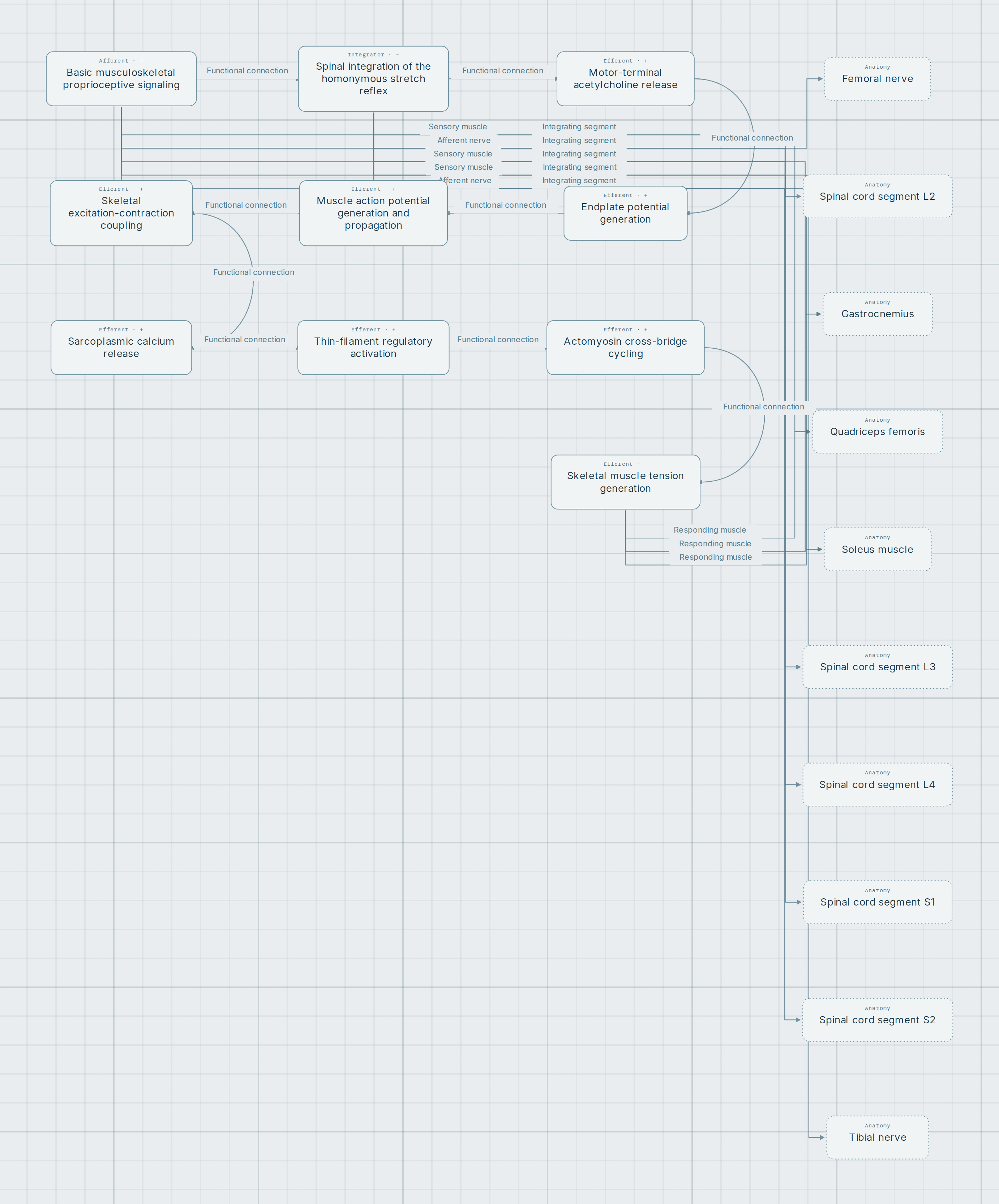
What the arrows mean
Follow the arrows, rather than just the boxes: the compact path changes direction between rows. The map combines equivalent processes for comparison. Notes retain the reflex and conditions belonging to each application.
- Functional connection
- Orders circuit components. It does not assign a numerical sign or imply that every box represents a neuron or synapse.
- Sensor muscle and afferent nerve
- Locate the sensory input. Calling a muscle a sensor here refers to its spindles; it does not turn the entire muscle mass into a receptor.
- Integrating segment and response muscle
- Locate integration and the effector. Segments do not necessarily contribute equally, and a branch does not diagnose a lesion.
From sensory input to response
Input and spinal core
Stretch activates muscle spindle afferents. The homonymous excitatory core connects Ia afferents with alpha motor neurons; reciprocal inhibition requires a separate interneuronal organization that is not expanded here. The integration node summarizes the function rather than representing every neuron.
Two anatomical maps
The patellar application connects quadriceps, femoral nerve and L2–L4 segments. The Achilles application connects gastrocnemius and soleus, tibial nerve and S1–S2. These representative assignments remain attached to their circuit: a shared “spinal integration” process does not merge their levels or peripheral pathways.
From electrical signal to force
The motor-response branch proceeds through acetylcholine release, end-plate potential and muscle action potential. It then shows excitation–contraction coupling, calcium release, thin-filament regulation, cross-bridge cycling and tension. This extension explains why the outcome also depends on the neuromuscular junction and muscle.
Monosynaptic does not mean one step to contraction
The term describes the excitatory spinal Ia–alpha motor neuron core. It does not remove sensory transduction, axonal propagation, neuromuscular transmission or contractile stages. Nor does it describe every circuit that modulates the response as monosynaptic.
The end-plate potential is a graded local depolarization. A propagated muscle signal requires threshold to be reached in an excitable membrane. The map separates these processes before continuing to reticular calcium release and actin–myosin interaction. It is not a measurement of latency or amplitude.
What the comparison shows, and what remains outside it
Combining the reflexes exposes mechanisms that recur in both. Expanding their locations reveals anatomical differences. The conditions specify homonymous laterality where applicable, but this snapshot does not fix a right or left side for these two reflexes.
An observed response also depends on posture, load, descending modulation and neuromuscular state. This guide therefore organizes anatomical and physiological reasoning; it does not assign a cause to a diminished, brisk or absent reflex or replace a complete examination.
A shared process does not permit a jump between circuits
A common node summarizes a reused function. To follow one reflex, retain its identity when reading muscles, nerves, segments and conditions. Visual merging is not an additional functional connection.
Explore it yourself
- Follow the nine visible connections in the main figure, separating neural signals, neuromuscular transmission and contraction.
- Open the anatomical expansion and select spinal integration. Compare the patellar and Achilles notes before interpreting segmental levels.
- Deselect one reflex in Filters and navigation. Observe which applications disappear and which sequence remains.
Opens both reflexes together. Click a component to expand its anatomical sites; the notes separate each circuit’s applications.
Scope and sources
A representative physiological map, not a patient record. Reflex connections retain partial support; they do not express equal segmental contributions or a complete central circuit. The anatomical figure expands only three components. The side selector does not impose laterality on these reflexes, whose side is unspecified in this snapshot.
References recorded in the KG applications and complementary teaching material. They explain the circuit or mechanism without making this view a diagnostic algorithm:
- Purves and colleagues. Neuroscience, 2nd ed. The Spinal Cord Circuitry Underlying Muscle Stretch Reflexes. NCBI Bookshelf: Ia–alpha core and associated circuits.
- Walker, Hall and Hurst, editors. Clinical Methods, 3rd ed. Deep Tendon Reflexes. NCBI Bookshelf: examination principles and contextual interpretation.
- OpenStax/Rice. Anatomy and Physiology 2e, 14.3. Motor Responses: somatic reflex organization, including excitatory and inhibitory branches.
- The excitation-contraction coupling mechanism in skeletal muscle. 2014; PMID 28509964. Mechanistic review: CaV1.1, RyR1 and calcium release, not a reflex segment map.