Anatomy · Lymphatic networks
Lymphatic drainage: different territories, shared pathways
A lymph node group is understood through more than its name or location. Its incoming territories, shared pathways and possible outlets also matter. This map compares two axillary inputs and follows their common connections toward downstream routes while preserving the direction of drainage.
Explore this map in the AtlasOpens pectoral and humeral groups, their central and apical connections, and the pectoral territories. Expand the outlets afterward.
Two scales of one pathway
Expansion: cervical pathways and venous outlets


Reading inputs and outputs
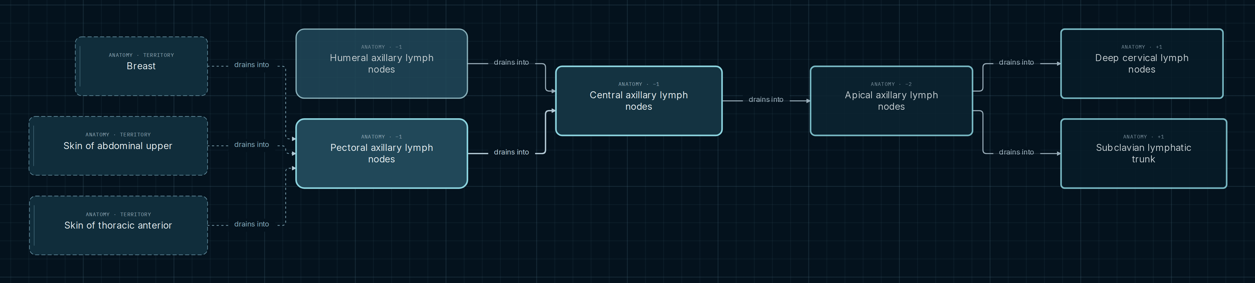
Start with the territories on the left and follow each arrow toward the receiving group. Compare the pectoral and humeral groups: they have their own inputs but share part of the displayed pathway. Selecting a node exposes its territorial inputs; changing selection does not mean the previous territories have stopped draining.
- Drains into
- Connects an origin to a recorded drainage recipient. The arrow stays unchanged when you explore upstream inputs.
- Convergence
- Two groups point to the same canonical node. A shared segment can be recognized without duplicating it in two independent lists.
- Alternative outlet
- The apical connection to deep cervical nodes accompanies the route to the subclavian trunk. It is not a sequence from apical to cervical to subclavian nodes.
- Territory and location
- A structure draining into a group is not necessarily the region where that group is located. These are distinct relationships.
Where integration becomes visible
Territorial inputs
The pectoral selection shows Breast and the skin of the anterior thoracic and upper abdominal walls. The breast note limits the relationship to an anterolateral route, not the entire breast. Skin routes are superficial and nonexclusive. Node size does not represent the amount of lymph carried.
A shared segment
Pectoral and humeral groups point to central nodes, which continue toward apical nodes. Comparing them shows which connections are specific and which remain when one group is removed from the selection. This subset does not exhaust all axillary connections.
Two outlets from apical nodes
The map records an outlet toward the subclavian lymphatic trunk and an additional connection to deep cervical nodes. The second figure continues the cervical route to the jugular trunk and shows ductal alternatives with side qualifiers.
What comparing pathways reveals
A list makes each structure look independent. A network shows a central node receiving connections from different inputs. This supports organizational questions: which part changes when another group is selected, where does a common segment begin, and which destination requires a laterality note? These questions are answered by following relationships rather than accumulating names.
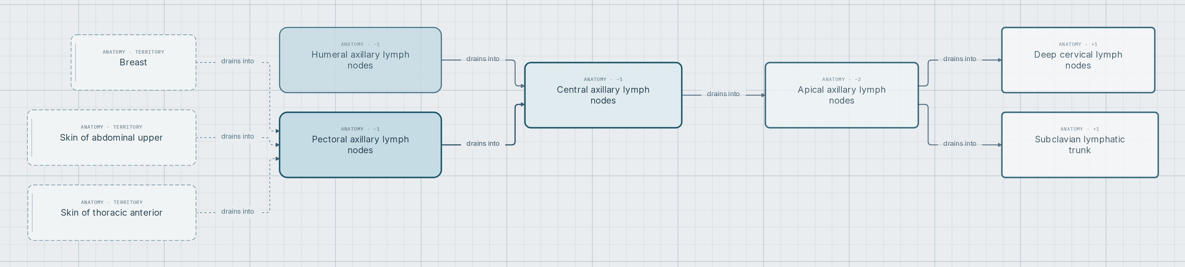
The initial selection stops before every outlet is expanded so the axillary comparison stays readable. The second figure extends the same pathway. These are different scales of reading, not separate anatomical networks.
Reading the end of the map
Jugular trunk connections to the thoracic and right lymphatic ducts carry side conditions. Notes also allow independent venous outlet alternatives. Displaying them together lets you compare patterns; it does not assert that one trunk in an individual patient follows both simultaneously.
A missing arrow in this selection does not establish anatomical absence. Open the Atlas to inspect additional connections and their context. Notes are part of reading the map: they explain what the drawing alone necessarily simplifies.
Sharing a node does not mean sharing all its conditions
A common identity helps compare pathways. Anatomical interpretation still requires the side, territorial scope and outlet variants. Do not trace a route across left and right simply because both use the same structure name.
Explore and compare
- Follow the three territories toward pectoral nodes and find where their pathway joins the humeral route.
- Open the map and click the humeral group to change the highlighted territories. Compare the new input with the central segment that remains.
- Expand toward the outlets. At the jugular trunk branches, examine the side qualifiers and venous outlet notes.
- Switch to Inputs to explore upstream. The exploration direction changes, but arrows still represent recorded anatomical drainage.
Opens pectoral and humeral groups, their central and apical connections, and the pectoral territories. Expand the outlets afterward.
Scope and sources
Selective macroscopic drainage view. It does not represent quantitative flow, an individual lymphogram, a sentinel node study or tumor spread. The Subclavian vein recipient simplifies an outlet whose precise site is described in notes as the venous angle; the figure is not vascular geometry. Explicit regional-location coverage for lymph node groups is incomplete. The upper abdominal wall skin connection is recorded in the KG; the linked institutional table does not explicitly name that territory in its pectoral-group description, so a specific supporting locator remains pending.
These institutional tables are the references associated in the Atlas with the groups and relationships examined. Relationship-specific locators and qualifications are retained; support for the abdominal territory needs the clarification noted above. Figures are screenshots of the selection, with no added pathways to complete an idealized tree.
- Texas Tech University Health Sciences Center El Paso · Lymphatics of the Thorax: axillary groups and outlets.
- Texas Tech University Health Sciences Center El Paso · Lymphatics of the Upper Limb: pectoral, lateral and central groups.
- Texas Tech University Health Sciences Center El Paso · Lymphatics of the Head and Neck: deep cervical nodes and jugular trunk.