Physiology · Digestive system
Gastric acid secretion: converging signals in the parietal cell
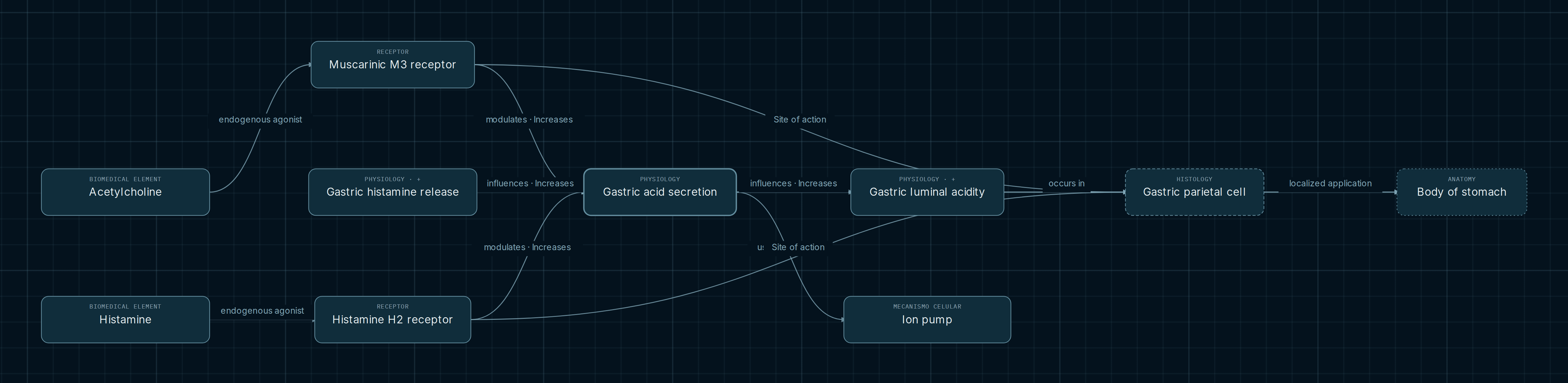
Two signals can regulate the same function without being equivalent. This map brings acetylcholine/M3 and histamine/H2 together around gastric acid secretion, linking that function to the parietal cell, an ion transport mechanism class and luminal acidity. Their convergence helps separate the contribution of each explanatory level.
Explore this map in the AtlasOpens acid secretion with receptors, mechanism, histology and anatomy.
The map
What each connection means
Read the ligand–receptor pairs first, then their arrows toward secretion. “Site of action” places both receptors in the parietal application. “Effector participant” connects that cell to the function. Anatomy locates the system; it does not add a signaling step between receptor and secretion.
- Endogenous agonist
- Identifies a substance capable of activating the subtype. Acetylcholine does not act exclusively at M3, nor does histamine act exclusively at H2.
- Modulates · increases
- Indicates a stimulatory influence with preserved activation and coupling. Each edge expresses an effect; it does not calculate the net response to all signals.
- Uses mechanism and occurs in
- Ion pump supplies a mechanism class; gastric body supplies a representative location. Neither relationship alone is equivalent to the complete function.
Signals and their shared function
Two receptor inputs
Acetylcholine/M3 and histamine/H2 reach the same secretory function and parietal context. Convergence indicates a shared functional target without implying equal potency, identical intracellular mechanisms or a linear sum of responses. The M3 application retains its partial-support designation.
Histamine release
Gastric histamine release is a process; histamine is the chemical entity that activates H2. The map connects release to acid secretion through a stimulatory influence. Notes describe a paracrine signal from ECL cells, although those cells are not displayed as a node in this selection.
Effector and location
The parietal cell represents the oxyntic phenotype in fundic/body glands. Its gastric body application does not assign acid secretion to the entire epithelium or exclude other territories. Histological context defines where receptor and function links should be interpreted.
From transport mechanism to luminal outcome
The “Ion pump” node is a general cellular function class. Its relationship note mentions primary H/K transport as a contribution to secretion, but this view does not include a specific molecular pump. Receptor and pump have different roles: one receives a regulatory signal, while the transport mechanism contributes to ion movement.
Acid secretion and increasing luminal acidity are also different concepts. Their relationship holds buffering capacity and gastric emptying constant. If those conditions change, the same secretion can accompany a different acidity. Greater acidity means lower pH; the “increases” arrow refers to acidity rather than to a numerical increase in pH.
What each source demonstrates
The human study by Pfeiffer and colleagues characterizes M3 receptors through radioligand binding in gastric mucosa. That design supports the subtype in the tissue, but does not independently identify every receptor-bearing cell or measure secretion by an isolated parietal cell. The parietal application therefore combines sources and retains its partial-support classification.
Tanaka-Shintani and Watanabe examine immunostaining and cellular distribution in resected stomachs. Their work provides context for the ECL population; detecting markers is not equivalent to demonstrating histamine release. IUPHAR catalogs clarify receptor identities and ligands, while physiological texts contextualize the integrated function. These are complementary contributions with different evidential scopes.
Convergence means a shared function, not a calculated net response
Two stimulatory arrows are insufficient to calculate acid output. The notes require viable cells and functional machinery, while acknowledging other concurrent signals. The map organizes selected influences; it does not include an integration equation.
Explore for yourself
- Follow each ligand to its receptor and compare where M3 and H2 converge. Open the notes for both modulatory relationships.
- Inspect the parietal cell and its gastric body application. Distinguish site of action, effector participant and anatomical location.
- Follow secretion toward luminal acidity and read the buffering and emptying conditions. Compare the human tissue, histological and regulatory references. Some notes remain in their original Spanish.
Opens acid secretion with receptors, mechanism, histology and anatomy.
Scope and sources
The selection does not represent every gastric regulator, inhibitor or feedback circuit. It cannot establish a clinical response, a secretion magnitude or a treatment effect. It preserves distinctions between a function, a luminal variable, a mechanism class and a cellular context.
Sources recorded in the notes and checked for this guide. Human studies contribute specific evidence; the review is listed as contextual synthesis rather than an additional experiment:
- OpenStax/Rice. Anatomy and Physiology 2e, 23.4. Histology, secretory cells and gastric regulation.
- IUPHAR/BPS. Acetylcholine receptors (muscarinic), v2025.3. Abstract and M3 link; DOI: 10.2218/gtopdb/F2/2025.3.
- IUPHAR/BPS. Histamine receptors, v2025.3. Abstract and H2 link; DOI: 10.2218/gtopdb/F33/2025.3.
- Pfeiffer et al. Dig Dis Sci. 1990;35:1468–1472. PMID: 2253531. Radioligand binding in human gastric mucosa.
- Tanaka-Shintani and Watanabe. Pathol Int. 2007;57:572–583. PMID: 17685928. ECL immunohistochemistry in resected human tissue.
- Engevik, Kaji and Goldenring. Physiol Rev. 2020;100:573–602. PMID: 31670611. Parietal physiology review; figure 4: receptors.
- Alberts et al. Molecular Biology of the Cell. Carrier Proteins and Active Membrane Transport. NCBI Bookshelf: NBK26896.