Pathophysiology · Liver and circulation
Cirrhosis: contributors, mechanisms and consequences
Cirrhosis brings together a history of liver injury and consequences that can involve the circulation, kidneys and other organs. Studying it requires distinguishing diseases that may precede it, pathophysiological changes and clinical complications. This guide follows those three questions through relationships recorded in the Atlas.
Explore this map in the AtlasOpens four selected states and their immediate connections: fibrosis, portal pressure, sinusoidal pressure and ascites.
The map
Converging diseases and diverging complications
How to read the connections
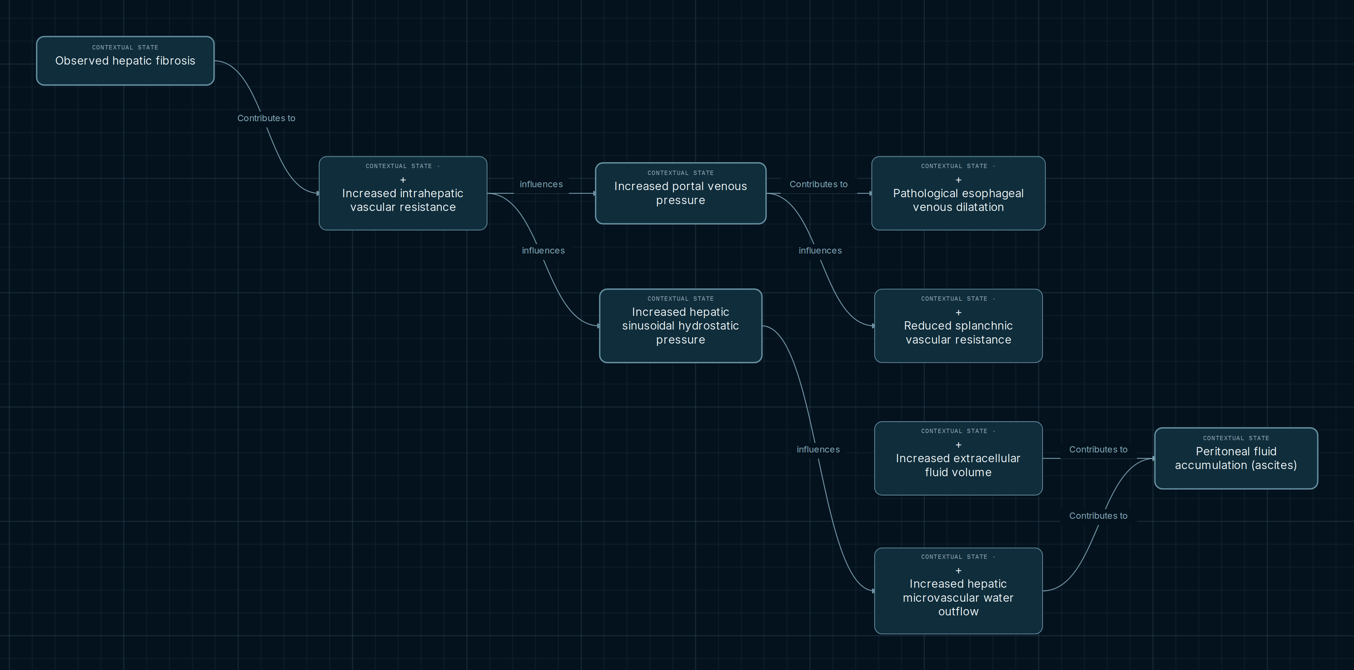
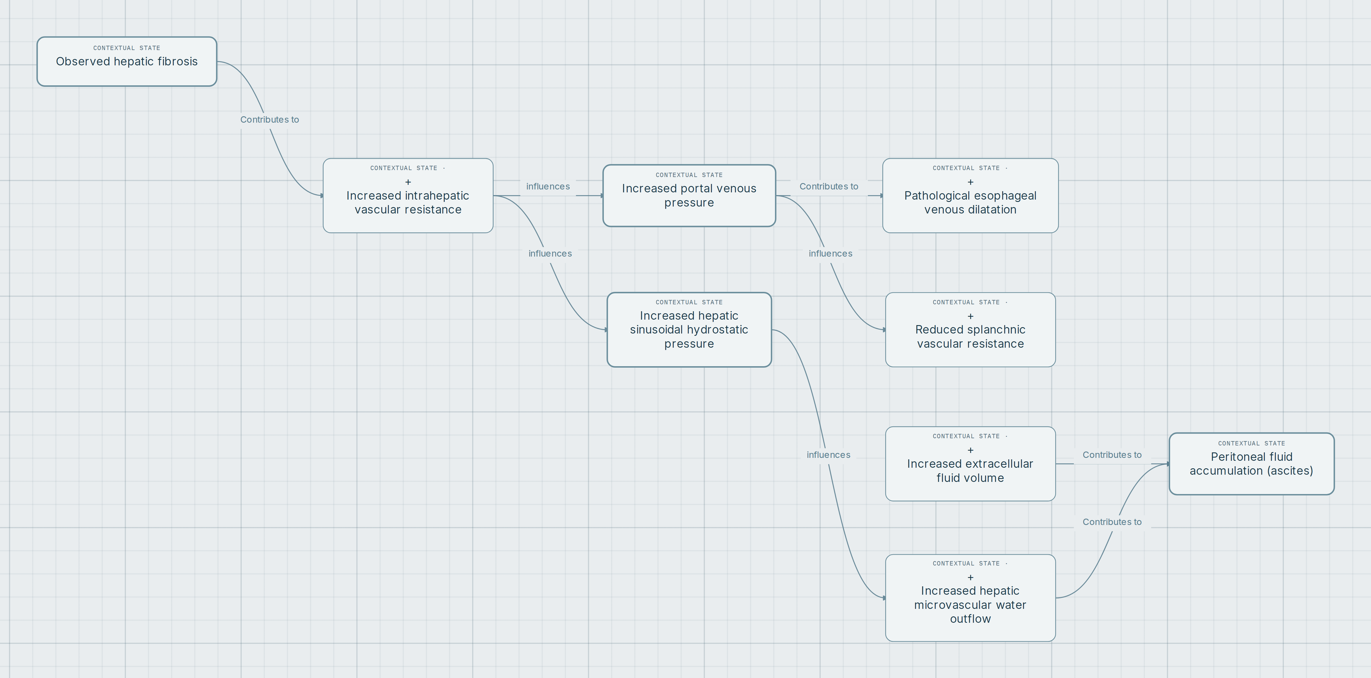
Start at fibrosis and follow intrahepatic resistance. Two branches emerge from that point and need to be read separately. Arrows retain the recorded relationship and its conditions. The selection brings observed states together; it does not depict an inevitable individual course or calculate the net effect of several influences.
- Contributes to
- Indicates a conditional contribution. Fibrosis can contribute to higher resistance when vascular architecture is distorted; the relationship does not establish sufficiency.
- Influences
- Relates variables within a documented context. In the vascular branch, flow, the location of resistance and downstream pressure must be considered.
- May be complicated by
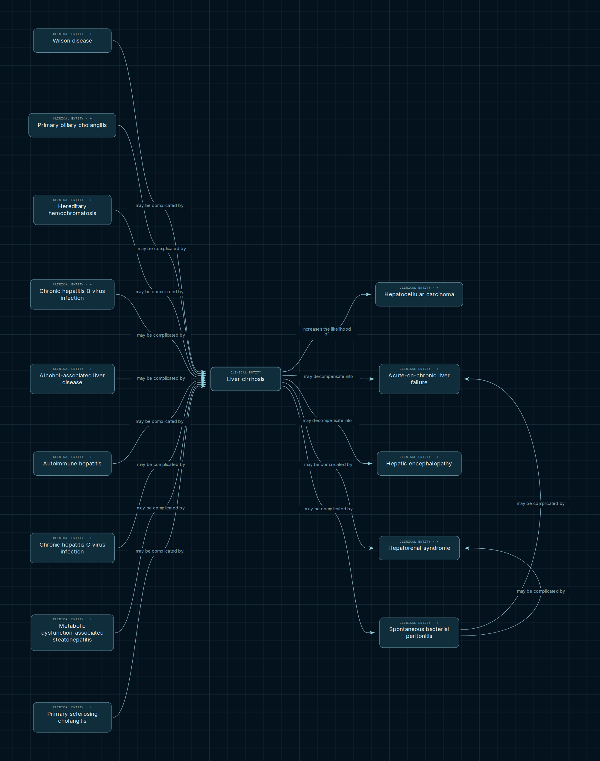
- In the clinical view, this expresses a possibility between entities. A possible complication is not a required stage or an individual prediction.
Three points along the pathway
Fibrosis and resistance
The starting node describes observed hepatic fibrosis. Its note requires excessive fibrous matrix in the liver and distinguishes that finding from a diagnosis or stage. The following connection requires demonstrated increased intrahepatic resistance.
Two pressures, two pathways
Portal pressure connects with pathological dilation of esophageal veins and reduced splanchnic resistance. Sinusoidal pressure connects with increased hepatic microvascular water outflow. These are different vascular compartments, each with its own conditions.
Convergence on ascites
The map retains two contributors to peritoneal fluid accumulation: increased hepatic water outflow and increased extracellular volume. This convergence helps compare distinct contributions without attributing every instance of ascites to one mechanism. Notes distinguish movement from blood into interstitium from subsequent delivery into the cavity: the latter contributes when it exceeds peritoneal or lymphatic removal.
What can contribute to cirrhosis
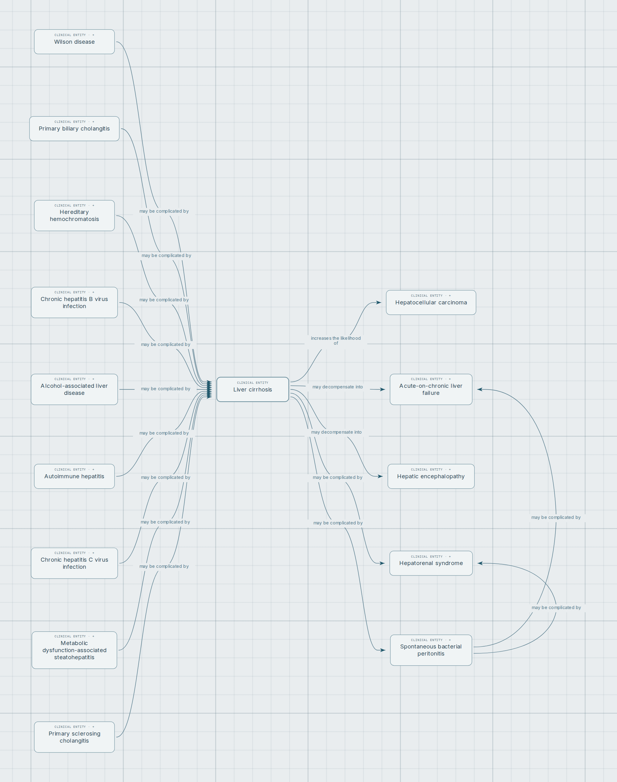
The clinical view records incoming relationships from chronic hepatitis B, chronic hepatitis C, alcohol-associated liver disease and metabolic dysfunction-associated steatohepatitis. It also includes branches involving autoimmune hepatitis, hereditary hemochromatosis and biliary disease. Each relationship identifies the relevant branch of progressive injury; it does not turn every infection, exposure, instance of steatosis or genetic carrier state into cirrhosis.
A useful study exercise is to compare two incoming branches: identify the disease represented by each node, read the arrow label and inspect the condition limiting its scope. Sharing a destination does not mean sharing every mechanism or following the same timeline.
Which clinical consequences are recorded
Outgoing connections include hepatic encephalopathy, hepatorenal syndrome, spontaneous bacterial peritonitis and hepatocellular carcinoma. Their operators distinguish decompensation, complication and increased risk. Notes restrict, for example, the hepatorenal branch to its relevant setting and the peritoneal infection branch to spontaneous infection of ascites without a secondary abdominal source.
These clinical connections retain their own supporting evidence. The hemodynamic pathway in the main image should not be presented as a complete explanation for each one. An arrow also supplies no numerical probability, uniform time interval or diagnostic criteria evaluated by the map.
Observed fibrosis and clinical cirrhosis are distinct identities
The pathophysiology view begins with a tissue finding. The clinical view selects the entity “Liver cirrhosis.” Comparing them changes the level of explanation, but does not justify drawing an equivalence or an unrecorded causal bridge between them.
Explore for yourself
- Follow fibrosis → intrahepatic resistance and compare the notes for portal and sinusoidal pressures. Pay particular attention to the conditions describing vascular location.
- Select ascites and open “Notes and legend.” Compare its two incoming connections and inspect the attached references.
- In Clinical entities → Digestive, select Liver cirrhosis, both directions and depth one. Compare incoming and outgoing relationships with the pathophysiology map. Some source notes remain in Spanish.
Opens four selected states and their immediate connections: fibrosis, portal pressure, sinusoidal pressure and ascites.
Scope and sources
This selection shows part of the published pathophysiology and clinical relationships. It does not enumerate every etiology, manifestation or complication. It does not include the complete renal retention pathway here or allow sinusoidal pressure to be inferred from presinusoidal portal hypertension. The map is educational and does not assess individual cases.
Atlas notes preserve references and locators for individual connections. The sources let readers review the general context separately from the mechanism represented:
- AASLD. Back to Basics: Outpatient Management of Cirrhosis (2024), hemodynamic introduction and Figure 1.
- NIDDK. Cirrhosis, definition and overview of causes.
- Hepatic Circulation — Fluid Exchange, ascites formation section; reference recorded in the graph.
- AASLD. Why do we use albumin in patients with cirrhosis? (2020), discussion of effective arterial volume.
- CDC. Clinical Overview of Viral Hepatitis (2025), consequences of chronic hepatitis B and C infection.
- AASLD. Diagnosis, Evaluation and Management of Ascites, Spontaneous Bacterial Peritonitis and Hepatorenal Syndrome, 2021 guidance portal.