Physiology · Cardiovascular
Cardiac output: why heart rate is only part of the story
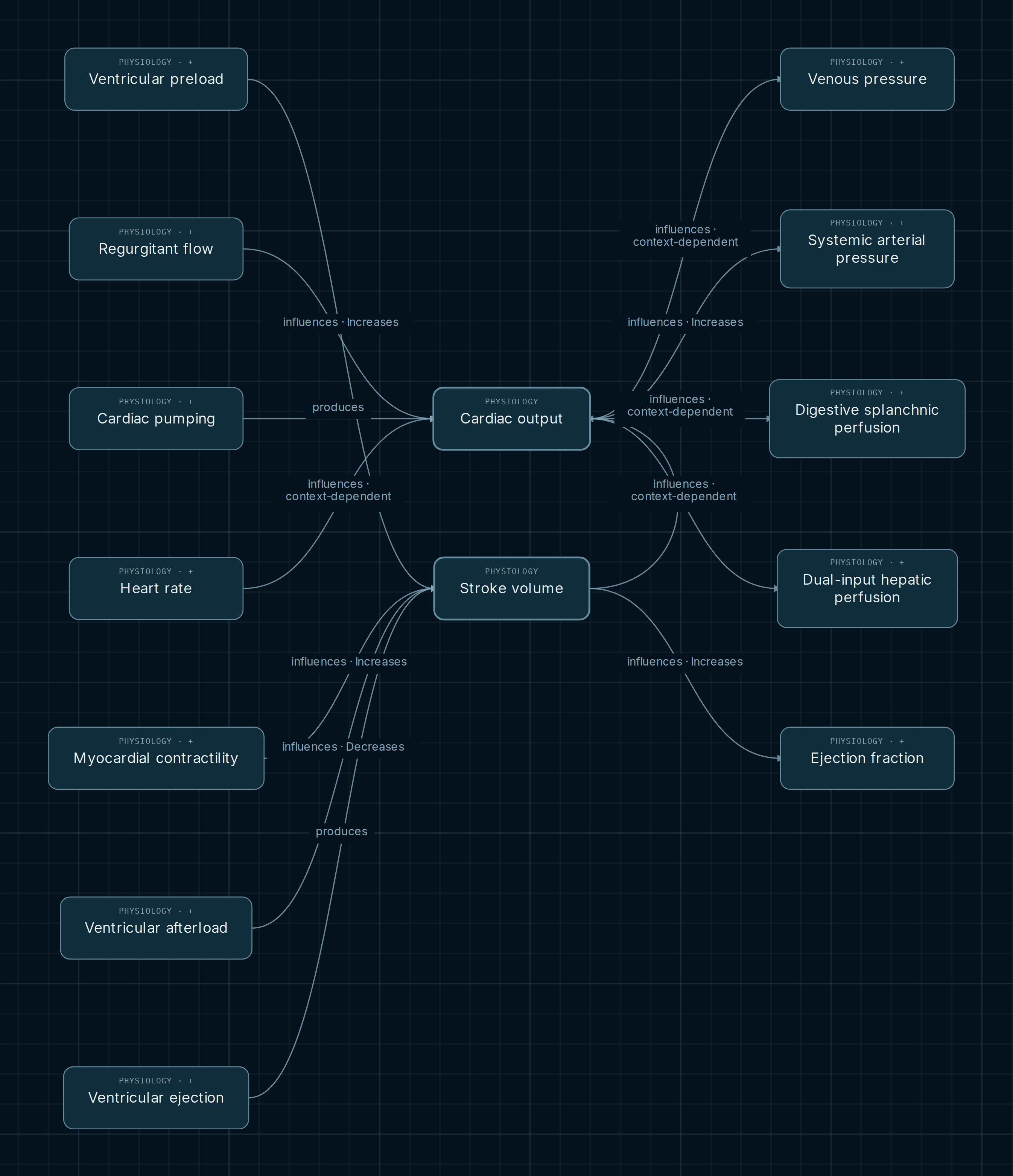
Cardiac output relates the volume ejected per beat to the number of beats per minute. The formula is short; its regulation is not. Changing heart rate can alter filling, and the same ejected volume does not always represent the same forward flow. These two views trace the determinants while retaining the conditions attached to each influence.
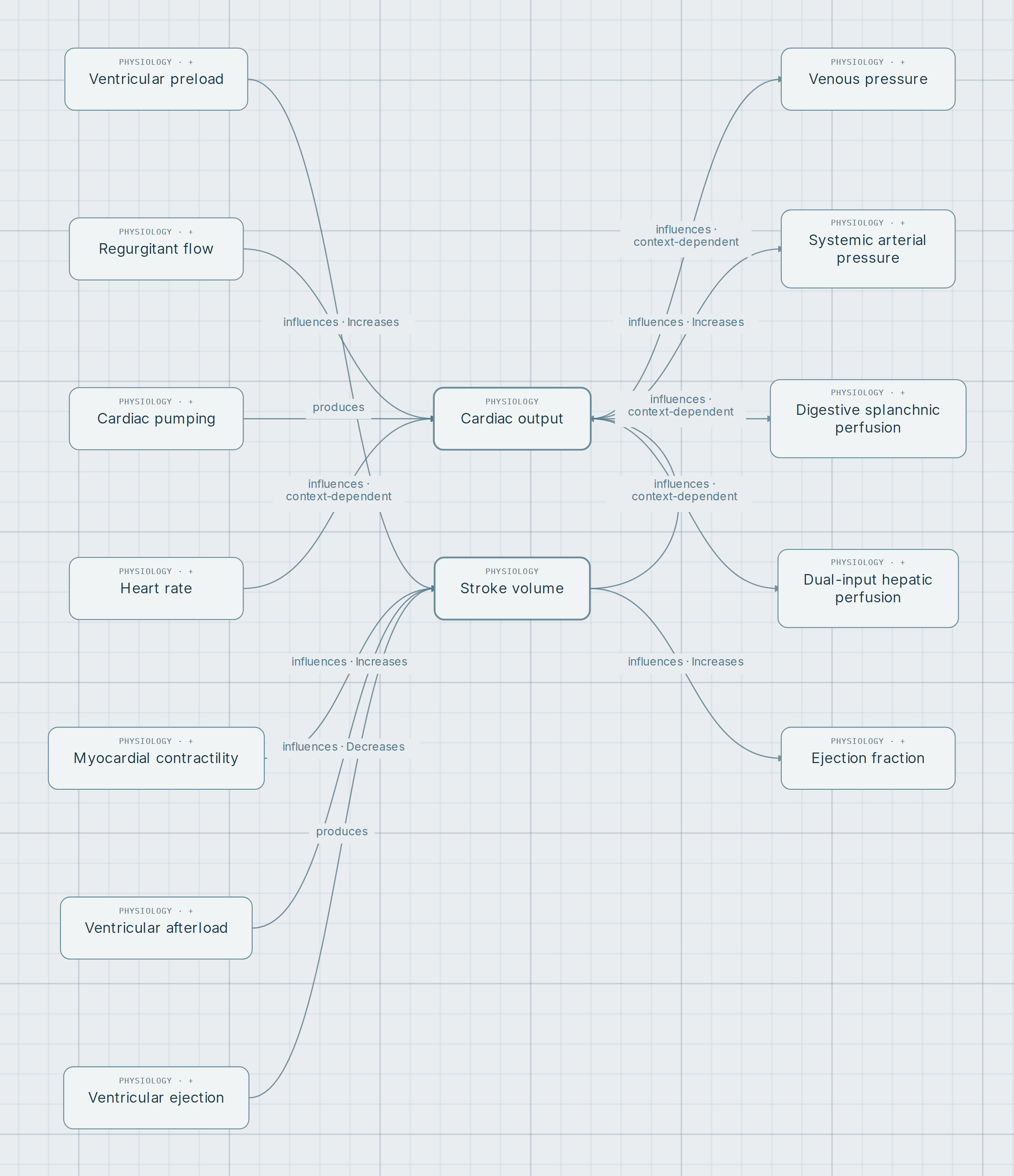
Explore this map in the AtlasOpens cardiac output and stroke volume with their immediate connections. The second figure separately explores heart-rate regulation.
Two complementary maps
Heart rate: mediators, receptors and context
How to read an influence
Start at stroke volume and cardiac output. Compare their inputs before following outputs toward pressure or perfusion. “Increases” describes an influence under stated conditions; “context-dependent” requires examining what can change. Neither label allows signs to be multiplied throughout every branch.
- Produces
- Cardiac pumping → output and ventricular ejection → stroke volume express process outcomes. Output refers to each ventricle, not their sum.
- Influences: increases or decreases
- Preload and contractility favor stroke volume under specific conditions. Increased afterload can reduce it in an acute change with other variables held constant.
- Modulates / context-dependent
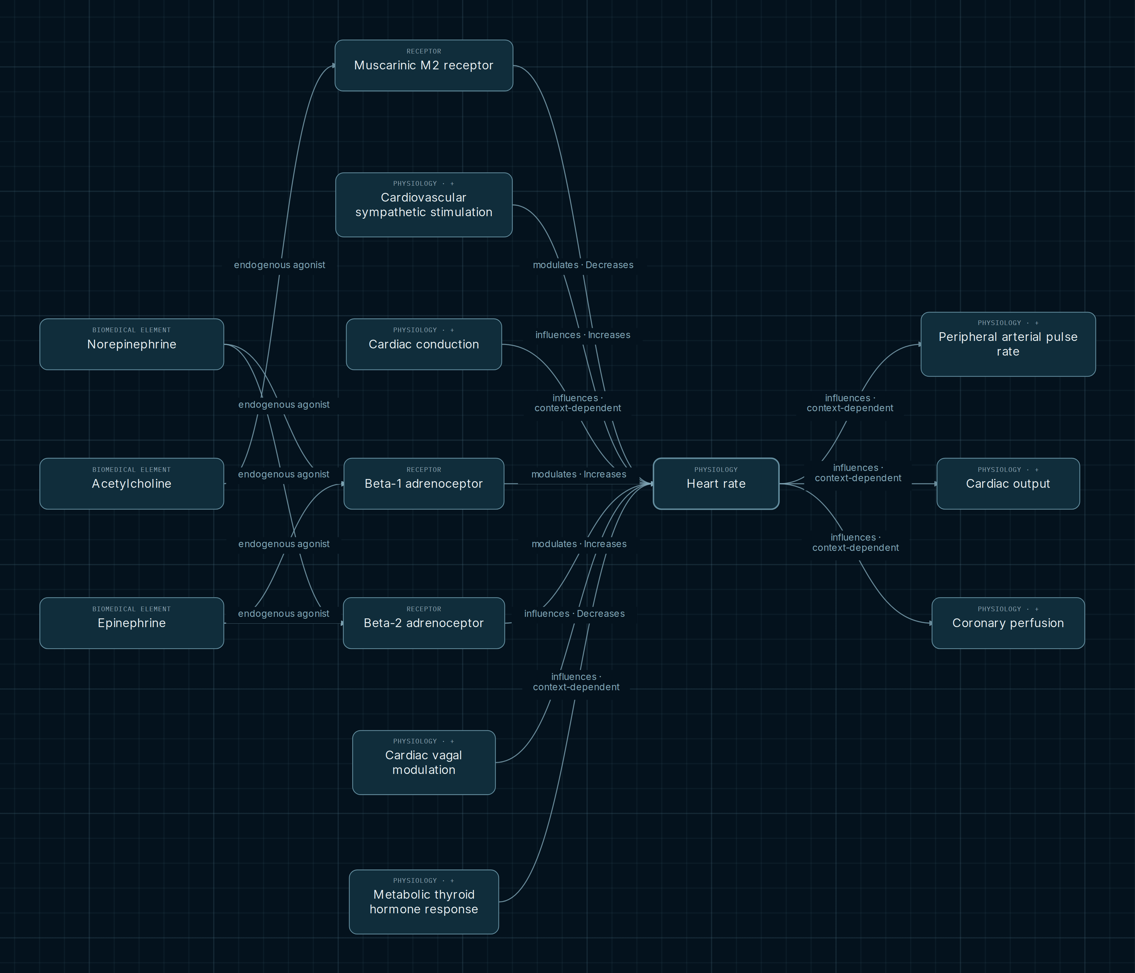
- The heart-rate view separates receptor activation from systemic influences. Effects on output, coronary perfusion or pulse cannot be resolved through one universal rule.
Determinants worth separating
A useful identity, not a guaranteed response
Cardiac output = heart rate × stroke volume, using consistent flow definitions. At fixed stroke volume, increasing rate raises output. In the body, volume per beat can change: tachycardia can limit filling. The heart rate → output connection retains exactly this qualification.
Preload, afterload and contractility
Three inputs converge on stroke volume. Preload acts on the ascending physiological portion with comparable loading and inotropic conditions. Contractility increases the capacity for ejection at comparable loads. They are not two names for one mechanism; the notes avoid equating Frank–Starling with increased contractility.
Ejected volume and effective forward flow
The regurgitant-flow connection uses a specific mitral context: at comparable total ejected volume and rate, a larger backward fraction reduces net systemic output. Compensation can change the result. Two stroke-volume values cannot be compared meaningfully without knowing which flow they represent.
Receptor regulation is not a sum of arrows
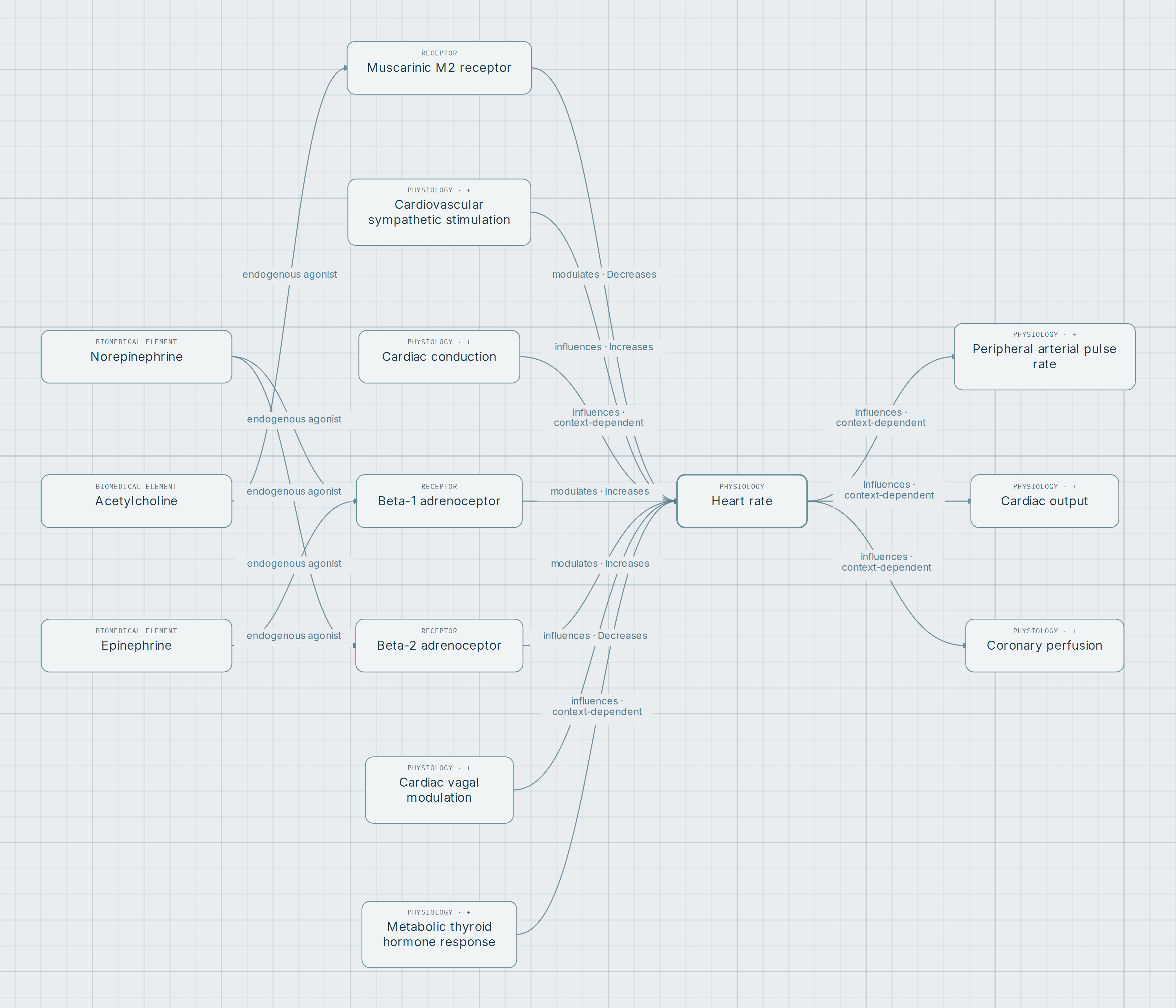
The second figure shows β1, β2 and M2 alongside their mediators. β2 notes retain human cardiac evidence that depends on agonist and region. M2 places the cholinergic effect in a functioning sinus-node context. These are conditional contributions, not an automatic prediction of final heart rate.
Sympathetic stimulation and receptor activation can describe different levels of the same response. Counting both branches as independent stimuli would duplicate their interpretation. The view compares levels of explanation; it does not add their effects or include all cardiovascular feedback.
Output, pressure, perfusion and ejection fraction
Higher cardiac output favors arterial pressure if systemic resistance and right atrial pressure remain fixed. It does not independently set each organ’s perfusion: hepatic and splanchnic branches retain resistance, redistribution and autoregulation conditions. The venous-pressure connection also does not imply that every low-output state causes congestion.
Ejection fraction is a proportion of end-diastolic volume; cardiac output is flow per unit time. The stroke volume → ejection fraction relation holds end-diastolic volume fixed. Sharing a determinant does not make these variables equivalent or make ejection fraction load-independent.
Before following an arrow, ask what must remain constant
The map is useful because it brings determinants together while preserving their conditions. The same local direction of influence can coexist with different overall responses when filling, load, rhythm or flow distribution changes.
Explore it yourself
- Compare the notes for preload, afterload and contractility. Identify what remains comparable in each relationship.
- Open the second figure and follow β1, β2 and M2 toward heart rate. Distinguish mediator, receptor and systemic influence.
- Review regurgitant flow, then outputs toward pressure, perfusion and ejection fraction. Find the conditions that prevent automatic conclusions.
Opens cardiac output and stroke volume with their immediate connections. The second figure separately explores heart-rate regulation.
Scope and sources
These are views of recorded relationships, not a hemodynamic simulation or dosing tool. No output, perfusion or pressure values are derived. Some relationships retain editorial-proposal status; the notes distinguish those scopes. The figures are complementary selections rather than the entirety of cardiovascular control.
KG references checked for this reading. Physiology sources explain conditional relationships; human research and receptor nomenclature support specific levels of the second view:
- OpenStax/Rice. Anatomy and Physiology 2e, 19.4. Cardiac output, heart rate, stroke volume and cardiac regulation.
- Klabunde. Effects of Preload, Afterload and Inotropy on Ventricular Pressure-Volume Loops. Independent effects with other variables held constant.
- Klabunde. Valvular Insufficiency (Regurgitation). Distinguishes total volume, regurgitant volume and forward flow.
- Klabunde. Central Venous Pressure. Volume, compliance and contributors to venous pressure, not a universal equivalence with output.
- Lemoine, Schönell and Kaumann. Br J Pharmacol. 1988;95:55–66. PMID 2851354. β1 and β2 receptors in human cardiac preparations.
- Birdsall and colleagues. IUPHAR/BPS Acetylcholine receptors (muscarinic), v2021.2. Nomenclature, acetylcholine and M2 coupling.