Anatomy · Nervous system
C5, C6 and C7 dermatomes: territories and shared regions
How do C5, C6 and C7 relate to regions of the upper limb? This map brings the three territories together so you can follow their connections, compare shared regions and read the notes attached to each relationship.
Explore this map in the AtlasOpens left-sided C5, C6 and C7 with their regions expanded.
The map
What each connection means
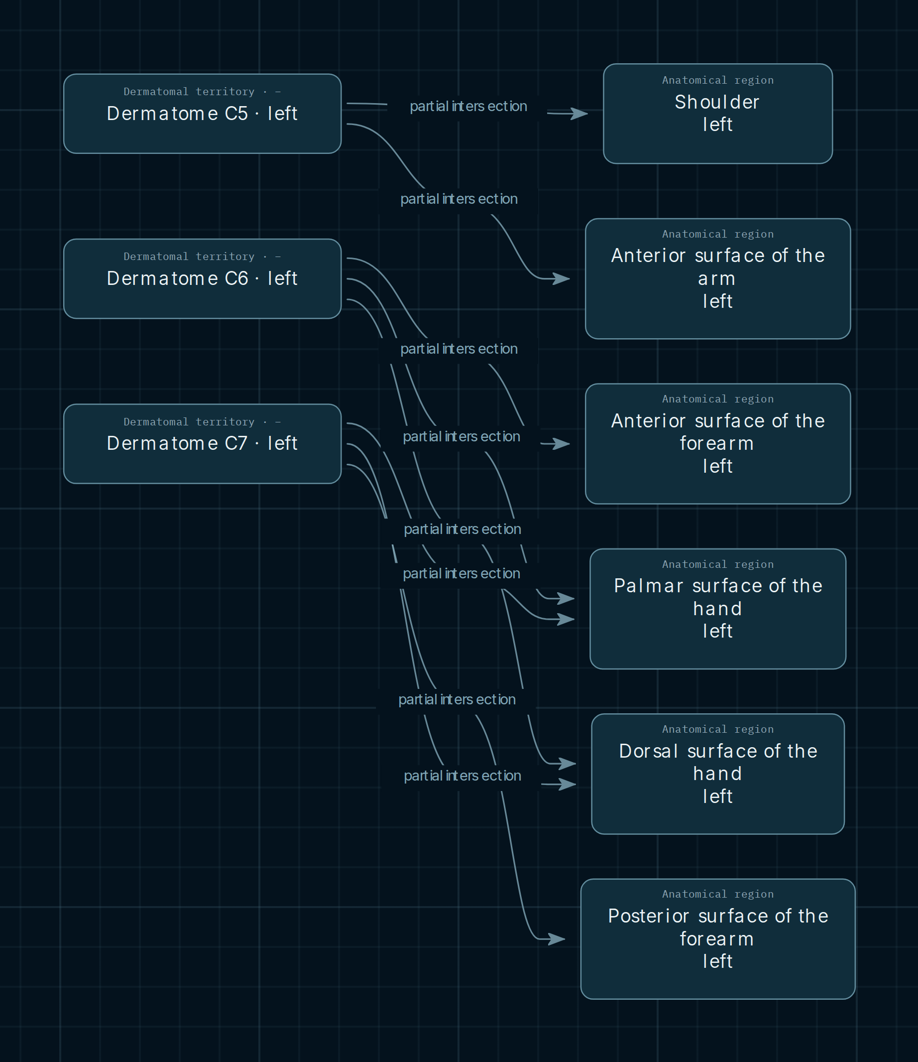
Dermatomal territories are on the left; the anatomical regions associated with them in this selection are on the right. Follow the arrows to compare those associations. They do not represent a nerve conduction sequence or a causal connection between regions.
- Partial intersection
- A dashed line connects a territory with a region it approximately intersects. It does not assign the entire region to that level.
- Shared region
- C6 and C7 converge on the palmar and dorsal surfaces of the hand. The shared node lets you compare associations; it does not mean they occupy identical points on the skin.
- Territory side
- The screenshot shows the left side. The Atlas selector lets you examine the right side separately, preserving connections for the selected side.
C5, C6 and C7 in this view
C5 · Shoulder and arm
The territory note describes the shoulder and lateral arm. The map connects it to Shoulder and Anterior surface of arm. These regional names are broader than the area described, so the relationship remains partial.
C6 · Forearm and hand
The description highlights the radial forearm and radial border of the hand. Its connections point to the anterior forearm and the palmar and dorsal hand surfaces. The diagram does not delineate digits or precise sensory boundaries.
C7 · Distal upper limb
The recorded description is the central distal area of the upper limb. It connects to the posterior forearm and to the palmar and dorsal hand surfaces. The latter are its points of convergence with C6 in this map.
A shared region is not an identical territory
The hand illustrates the distinction: two levels can relate to the same anatomical region without their sensory areas matching completely. The diagram shows regional associations; the notes clarify their scope.
Explore for yourself
- Open the map with all three territories expanded. Follow the lines from C6 and C7 to the hand and compare their forearm connections.
- Click C6 to close its connections, then open it again. Regions shared with C7 remain while that territory stays expanded.
- Select a territory or region and open “Notes and legend”. Review the description, relationship scope and source. Use “Territory side” to switch between left and right. Some source notes remain in Spanish and are identified as originals.
Opens left-sided C5, C6 and C7 with their regions expanded.
Scope and sources
A dermatome describes a skin territory associated with a spinal nerve level. This view represents approximate regional intersections, not precise boundaries. It is not a map of peripheral nerve territories or ISNCSCI examination points and cannot, by itself, attribute a symptom to a lesion. Evidence for the associations is recorded as partial.
The map refers to Lee, McPhee and Stringer (2008), figure 4, page 371. The Atlas retains this reference in the notes for territories and their connections. The image in this guide is an Atlas screenshot; the publication’s body map is available through the source: