Receptors · Histology · Anatomy
β1 and β2 receptors: from mediator to cell and organ
“β1: heart; β2: lung” is a useful memory aid, but it leaves out important biology. One mediator can activate both subtypes, one receptor can have several cellular contexts, and one cell can participate in different responses. This map helps you examine those overlaps and follow documented locations without turning a general association into a universal distribution.
Explore this map in the AtlasOpens β1 and β2 with their mediators and tissue applications. The search filter is ADRB1; β2 also remains visible because it is expanded.
The map
How to read the connections
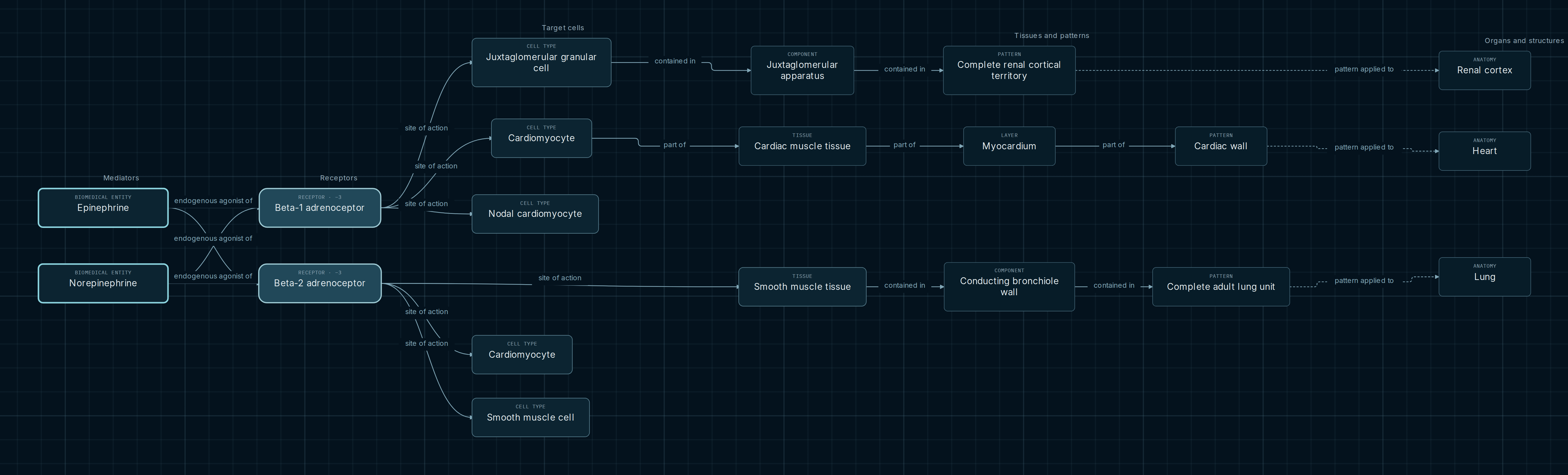
Read from left to right: mediator, receptor, site of action and, where an explicit assignment exists, tissue and organ. Arrows change meaning across columns. Receptor activation, tissue composition and the assignment of a histological pattern to a structure are different relationships.
- Endogenous agonist of
- Epinephrine and norepinephrine connect to both receptors. The four connections do not indicate equal affinity, potency or exclusive selectivity.
- Site of action
- Connects a receptor to a cellular or tissue context. Conditions distinguish working cardiomyocytes, nodal cells, vascular muscle and bronchiolar muscle.
- Part of / contained in / pattern applied to
- Trace a documented location. These are composition and assignment relationships, not steps in a signaling cascade.
Three contexts, one comparison
Heart: coexistence and different roles
β1 has working-cardiomyocyte and nodal-cardiomyocyte contexts. β2 also has a cardiomyocyte context. The application extending to the cardiac wall belongs to the working myocardium context of β1. A missing equivalent anatomical chain for the other contexts does not demonstrate that their receptors are absent from the heart.
Kidney: a specific cellular application
The renal β1 branch passes through the juxtaglomerular granular cell and juxtaglomerular apparatus to the cortical pattern. This is more informative than a generic association with “kidney”: it identifies a specific context of renin regulation. This receptor guide shows the location, without unfolding the entire renin–angiotensin sequence.
Airway: tissue and experimental scope
The bronchiolar β2 branch connects smooth muscle tissue, conducting bronchiolar wall, pulmonary pattern and lung. The vascular smooth muscle cell context remains separate. Sharing the words “smooth muscle” does not justify transferring a vascular response to the airway or all its regions.
A shared cell class is not an identical application
Cardiomyocytes occur in both β1 and β2 contexts. Lemoine and colleagues studied human atrial and ventricular preparations and the contribution of both subtypes to catecholamine responses. This coexistence challenges an exclusive receptor–organ pairing, but does not establish a universal functional proportion.
Two nodes labeled cardiomyocyte can appear in the diagram: one belongs to an occurrence in the cardiac wall, while the other retains a receptor context without that explicit anatomical application. The cell class is shared; the location evidence is not interchangeable. We therefore do not automatically extend both branches to the same organ.
Why a connection’s notes matter
The bronchiolar assignment retains partial support and an explicit regional extrapolation. Its experimental reference studied human airway smooth muscle cells, including their response after β2 desensitization. It is not an inventory of expression in every bronchiole of an individual.
This distinction is useful when studying pharmacology: a target and its tissue help frame questions about a response. Answering them still requires the agonist, its exposure and the signaling state. The map neither calculates a net effect nor converts a location into a treatment indication.
Location tells you where to ask; it does not predict the response
Follow each branch with its conditions. A shared mediator or cell class does not imply an identical functional contribution, or that every anatomical application is documented.
Explore it yourself
- Compare the four mediator connections and read their notes: they are not a ranking of potencies.
- Open the notes of both cardiomyocyte contexts. Distinguish cell class, histological application and anatomical structure.
- Follow the renal β1 and bronchiolar β2 branches. Check what is assigned and where the available information ends.
Opens β1 and β2 with their mediators and tissue applications. The search filter is ADRB1; β2 also remains visible because it is expanded.
Scope and sources
This is a selection of β1 and β2 contexts in the published KG, not an exhaustive expression atlas or a prescribing protocol. β3 and other receptors are outside its scope. Three contexts lack an explicit anatomical application in this snapshot; none is filled in by analogy. No relationships have been added to the KG.
Catalog references and editorial cross-checks. IUPHAR 2026.1 updates the nomenclature check; the KG retains its original 2023.1 reference. Experimental findings are used within the stated scope:
- Baker and colleagues. IUPHAR/BPS Adrenoceptors, v2026.1: nomenclature and endogenous agonists of beta subtypes.
- Lemoine, Schönell and Kaumann. Br J Pharmacol. 1988;95:55–66. PMID 2851354. Abstract of the study of β1/β2 receptors in human cardiac preparations.
- Kaur and Rout. Physiology, Renin Angiotensin System. NCBI Bookshelf/StatPearls: juxtaglomerular cells and sympathetic control; a monograph, not primary research.
- Epinephrine evokes shortening of human airway smooth muscle cells following β2 adrenergic receptor desensitization. AJP Lung, 2022. DOI: 10.1152/ajplung.00444.2021. Human cellular research, not a complete anatomical distribution.